biomolecules
Biomolecules are molecules that are vital for life processes. All living organisms require biomolecules to obtain energy, build cellular structures, carry out metabolic reactions, and store and transmit genetic information.
Organisms obtain energy and nutrients from food, which contains biomolecules in various forms. These biomolecules are broken down during digestion, absorbed into the bloodstream, and used by cells to maintain life.
monomers
Multiple monomers bond together chemically to form a polymer.
- Glucose molecules (monomers) form starch or glycogen (polymers)
- Amino acids (monomers) form proteins (polymers)
- Nucleotides (monomers) form DNA or RNA (polymers)
Organic Biomolecules
Organic biomolecules always contain carbon and hydrogen.. Most organic molecules are formed when multiple smaller molecules bond together.
The four main classes of organic biomolecules are:
- Carbohydrates
- Lipids
- Proteins
- Nucleic Acids
Macromolecules
Macromolecules are large organic biomolecules with a molecular mass greater than 1000 daltons (Da). They are typically polymers formed by the joining of many monomers.
Carbohydrates
Elements: Carbon, Hydrogen, Oxygen (CHO)
Carbohydrates are often found in the form of polymers and are broken down into simple sugars by enzymatic activity throughout the digestive process. For example, amylase in your saliva begins to break down carbohydrates into simple sugars which taste sweet- the more complex the carbohydrate is depends on the number of saccharide molecules and influences the body's ability to digest it into the simplest form. This is why if you eat something like bread you'll quickly get that sweet taste whilst stuff like beans taste less sweet (because beans contain a lot more complex carbohydrates than bread)
There are four types of carbohydrates, classified based on the number of saccharide molecules they have:
These have one saccharide molecule, they are the simplest form of carbohydrates. Some examples are Glucose, Fructose and Galactose
They have two saccharide molecules, this is where they become very vaguely complex but not really. Examples include Lactose and Maltose
1–10 saccharide molecules, now some teachers don't really teach this and instead just teach polysaccharides but oh well womp womp. Your example here is raffinose, often found in beans- it's a prebiotic and has 3 saccharide molecules
Now this is the most complex carbohydrate, it's the body's main form of storage because it is compact and can easily be broken down into energy. Examples include glycogen (how your body stores glucose), starch (how plants store sugars), cellulose, pectin and hemicellulose (types of fibre)
Lipids
Elements: Carbon, Hydrogen, Oxygen (CHO)
Lipids store long-term energy, form cell membranes, provide insulation, and play a role in hormone production. Lipids are hydrophobic, meaning they repel water and do not dissolve in aqueous solutions.
There are three types of lipids: Phospholipids, Triglycerides and Steroids
Triglycerides:
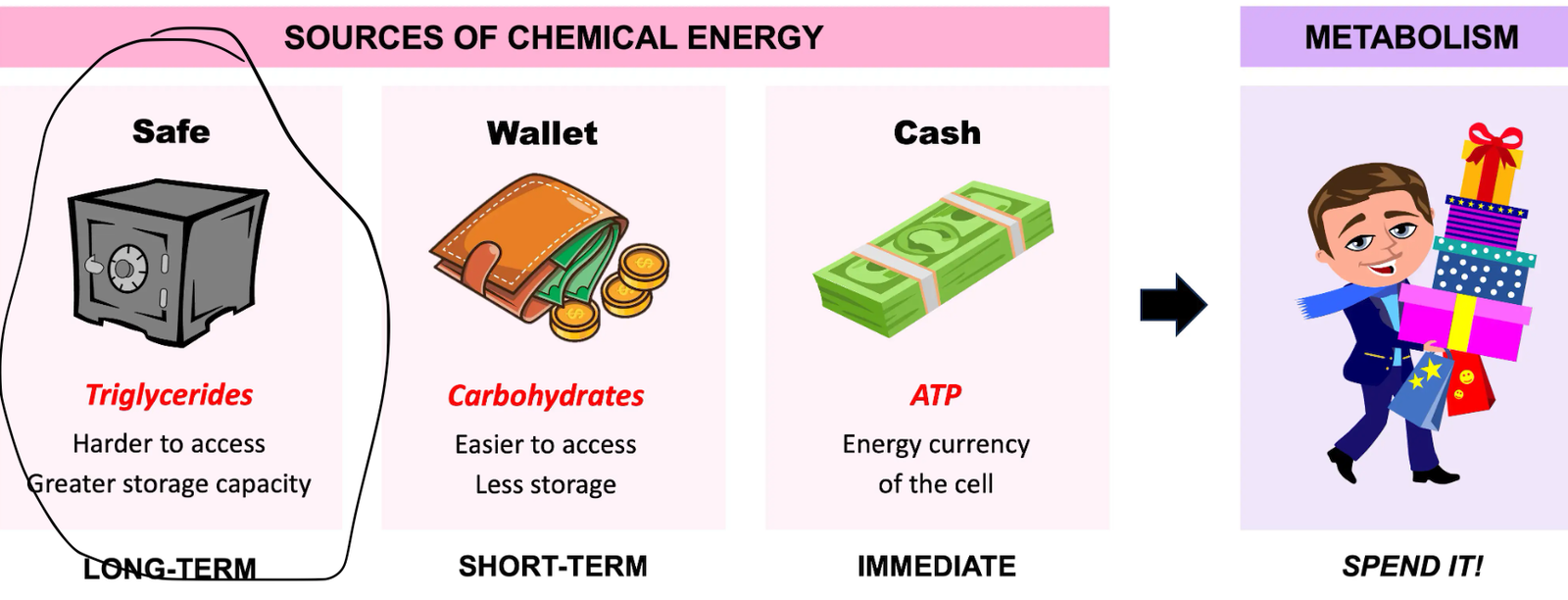
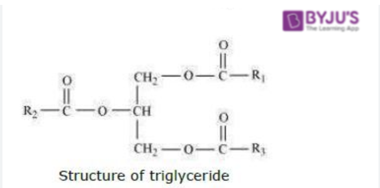
This is the most common form of lipid, consisting of three fatty acids and one glycerol backbone molecule. Triglycerides are a type of fat (lipid) found in your blood, essential for energy storage, but high levels can increase the risk of heart disease and other health issues.
Triglycerides are formed from the calories consumed for long lasting energy stores (your body doesn't immediately use these for energy)
Lipolysis is the breaking down of triglycerides into energy- this is done by Lipase separating fatty acids from the glycerol molecule
Phospholipids:
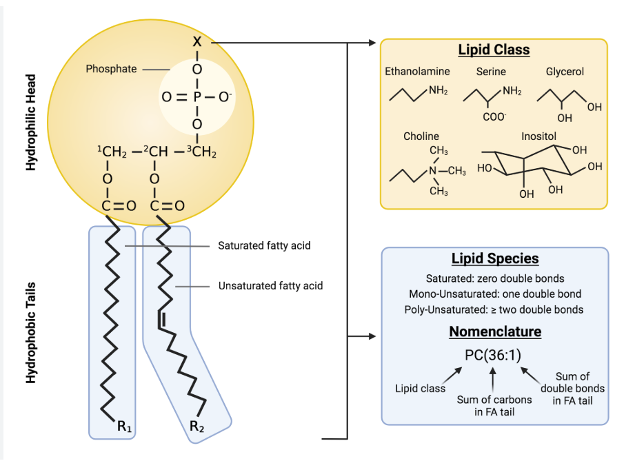
A phospholipid is a type of lipid molecule that is the main component of the cell membrane. Each phospholipid is made up of two fatty acids, a phosphate group, and a glycerol molecule. When many phospholipids line up, they form a double layer that is characteristic of all cell membranes.
Phospholipids are semi-permeable, this is an important characteristic as they are components of the cell membrane and therefore ensure only certain molecules can selectively pass through the sell membrane
Steroids:
The other type of lipids is steroids. In contrast to the phospholipids and triglycerides, steroids have a fused ring structure.
Although they do not resemble the other lipids, they are grouped with them because they are also hydrophobic and insoluble in water. All steroids have four linked carbon rings, and many of them, like cholesterol, have a short tail. Many steroids also have the –OH functional group, and these steroids are classified as alcohols called sterols.
Cholesterol is the most common steroid and is mainly synthesized in the liver; it is the precursor to vitamin D. Cholesterol is also a precursor to many important steroid hormones like estrogen, testosterone, and progesterone, which are secreted by the gonads and endocrine glands. Therefore, steroids play very important roles in the body’s reproductive system. Cholesterol also plays a role in synthesizing the steroid hormones aldosterone, which is used for osmoregulation, and cortisol, which plays a role in metabolism.
Excess cholesterol in the blood, also known as hypercholesterolemia, can lead to the buildup of plaque in arteries, increasing the risk of heart disease and stroke. This buildup can restrict blood flow and potentially cause chest pain or even heart attack or stroke if a clot forms. While high cholesterol often has no noticeable symptoms, it's crucial to monitor levels through blood tests and take steps to manage it through lifestyle changes and, if necessary, medication.
Fatty Acids
Fatty acids form the tail of the lipid molecule. There are two types of fatty acids: Saturated and Unsaturated: These are characterised by their cis carbon carbon double bonds- this means that they are unable to store molecules in a straight rod like shape, instead having a structural kink
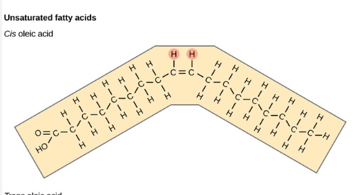
Saturated: These are the opposite of unsaturated, they don't have carbon carbon double bonds and store their molecules in a straight, rod like shape. These have a higher melting point compared to unsaturated fatty acids
Trans - Fats
Thought I'd mention it cause it's important too know in general. These are often seen as the unhealthiest fat, raising "bad fats"- low density lipids (LDLs) and lowering "good fats"- high density lipids (HDLs). Basically, trans fats especially in large amounts = bad, especially in criteria D essays. If you get something like this for an essay topic you can tie it into heart diseases like heart failure or atherosclerosis alongside how fast food contributes to this . In fact, here's a task idea: Write an essay in thirty minutes discussing the political, social, economic and cultural implications of the rise of fast food (I think it should be obvious to use trans fats and proper biological terminology in ur answer)
Which Is Healthier? Saturated or Unsaturated fats
Doctors generally recommend unsaturated fats as a healthier fat, this is because whilst saturated fats increase "good" and "bad" cholesterol (HDLs and LDLs); unsaturated fats are able to increase the good cholesterol whilst decreasing the bad cholesterol
It is generally recommended for 6% or under of an individual's diet to come from saturated fats and for 25% to come from unsaturated fats so do with that information what you will.
Saturated fats are usually solid at room temperature (butter for example) whilst unsaturated fats are liquid at this temperature (for example, olive oil)
Proteins
Elements: Carbon, Hydrogen, Oxygen, Nitrogen (CHON)
Proteins are crucial for a variety of functions, including muscle repair, making up your hair, skin and toenails- they also have a major role in the innate immune system as cytokines (which non-specifically attack an invading pathogen). The most abundant animal protein is collagen, which you might know is for your skin. Meanwhile, the most abundant plant protein is RuBiSCO
Proteins are fundamentally made out of peptides, now how do a bunch of random peptides form a protein you ask? PEPTIDE BONDS. That's actually all you've gotta know, now onto protein structures-
Protein Structures
Proteins must be folded multiple times to actually be useful to the body, there are four levels of folding before the protein becomes fully functional
- primary structure: This is like if you were tying your shoelaces, and you haven't started tying them yet. That makes the shoelaces kind of useless- likewise proteins in this stage are not folded at all and are hence useless or dysfunctional
- secondary structure: This structure would be like if you started tying your shoelaces but haven't finished yet- it doesn't work but it's closer to working that before ig
- tertiary structure: now this one would be like if you fully tied your shoelaces, it works finally!
- quartiary structure: now this is the final level of protein folding- it would be like if you double knotted your shoelaces in a sense
- prions: When proteins are misfolded they become a prion- this would kind of be like if you tied your shoes' laces together; it wouldn't work and you'd trip. Likewise, prions are uncurable and have a certain mortality rate- this is because not only are they completely dysfunctional but they also cause other proteins to misfold like them
Enzymes
Enzymes are biocatalysts that speed up cellular metabolic reactions by reducing the activation energy required for reactions to occur. They are made of proteins, are highly specific to their substrates, and can be reused repeatedly because they are not consumed during the reaction.
Enzyme activity is affected by several factors including temperature, pH, substrate concentration, and enzyme concentration.
Catalyst
A catalyst is a substance that increases the rate of a chemical reaction without being changed or used up by the reaction itself. Enzymes are biological catalysts.
Substrate
A substrate is the reactant molecule that binds to an enzyme. When the substrate binds to the enzyme, an enzyme–substrate complex is formed, which lowers the activation energy needed for the reaction to proceed.
Cellular Enzyme Types
Intracellular Enzymes
Intracellular enzymes function inside the cell. An example is enzymes involved in cellular respiration, which occur in the cytoplasm and mitochondria.
Extracellular Enzymes
Extracellular enzymes function outside the cell. Digestive enzymes such as amylase, protease, and lipase are secreted into the digestive tract to break down food molecules.
Nomenclature of Enzymes
Enzymes are usually named after the substrate they act on, with the suffix -ase.
- Lactose → Lactase
- Lipids → Lipase
- Proteins → Protease
Activation Energy
Activation energy is the minimum amount of energy required for a chemical reaction to occur. Enzymes lower this energy barrier, allowing reactions to happen faster and at normal biological temperatures.
Enzyme Action
Enzymes bind to their specific substrate to form an enzyme–substrate complex. This complex lowers activation energy and allows either a catabolic (breaking down) or anabolic (building up) reaction to occur, producing an end product.
Active Site
The active site is a specific region or groove on the enzyme where the substrate binds. It is divided into two functional regions:
- Binding site: The region where the substrate attaches to the enzyme.
- Catalytic site: The region where the chemical reaction takes place.
Lock-and-Key Model
The lock-and-key model explains enzyme specificity by stating that the shape of the enzyme’s active site is exactly complementary to the shape of its substrate. Only the correct substrate can fit into the active site, just as only the correct key fits into a lock. This explains why enzymes are highly specific.
Induced Fit Model
The induced fit model states that enzymes are specific but not rigid. When the substrate binds, the active site slightly changes shape to fit the substrate more tightly. This improves the interaction between enzyme and substrate and further lowers activation energy. However, enzymes cannot permanently change shape.
Factors Affecting the Rate of Enzyme-Controlled Reactions
Temperature
As temperature increases, kinetic energy increases, leading to more frequent collisions between enzyme and substrate and an increased rate of reaction.
- Approximately 40°C is the optimum temperature for many human enzymes.
- Above 40°C, enzymes undergo denaturalisation, causing the active site to lose its shape and the enzyme to stop functioning.
pH Level
Different enzymes have different optimum pH levels. As pH approaches the optimum, the rate of reaction increases.
- Deviations from the optimum pH cause changes in the enzyme’s structure.
- Extreme pH levels result in denaturalisation of the enzyme.The same goes for temperature
Substrate and Enzyme Concentration
Increasing substrate or enzyme concentration increases the rate of reaction because more enzyme–substrate complexes form.
- At a certain point, all active sites are occupied and the reaction rate plateaus.
Enzyme Use in the Food Industry
- Pectinase breaks down substances in apple cell walls, increasing juice extraction.
- Lactase breaks down lactose in milk, producing lactose-free milk.
- Proteases pre-digest proteins in baby food.
Other Uses of Enzymes
- Used in the treatment and prevention of diseases
- Involved in seed germination
- Used to produce low-calorie foods
Lactose Intolerance
Lactose intolerance occurs when an individual produces insufficient amounts of the enzyme lactase. As a result, lactose cannot be broken down into glucose and galactose, this means the lactose moves to the large intestine where it is fermented. This leads to bloating, gas discomfort and overall stomach pain. In order to manage this lactase can be added to milk to create lactose free milk (by breaking down the lactose into glucose and galactose, this version tastes sweeter). Additionally, a lactose intolerent individual can intake lactase supplements before consuming dairy to mitigate the side effects.
Structure of a Leaf
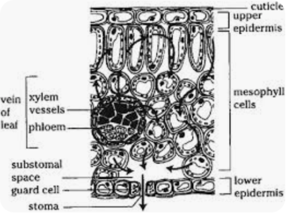
Picture taken from: ResearchGate
- Wax Cuticle: A protective, waterproof layer on the top of the leaf that prevents excessive water loss through evaporation.
- Upper Epidermis: A transparent layer that allows light to pass through to the palisade mesophyll layer underneath.
- Palisade Mesophyll: Tightly packed cells containing a large number of chloroplasts to maximize light absorption for photosynthesis.
- Spongy Mesophyll: Loosely packed cells with large internal air spaces, increasing the surface area for gas exchange.
- Lower Epidermis: Contains guard cells and stomata; this layer does not need direct light.
- Guard Cells: Specialized cells that absorb and lose water to open and close the stomata.
- Stomata: Openings where gas exchange (CO₂ and O₂) and water evaporation (transpiration) take place.
- Vascular Bundle: Contains xylem and phloem tissues.
- Xylem: Transports water and mineral ions from the roots into the leaf.
- Phloem: Transports sucrose and amino acids around the plant.
Chloroplasts
Chloroplasts are the organelles where photosynthesis takes place. A chloroplast consists of an outer membrane and an inner membrane, enclosing a fluid-filled region called the stroma.
Inside the chloroplast are flattened membrane-bound sacs called thylakoids. The thylakoids are arranged in vertical stacks known as grana (singular: granum), which are interconnected by lamellae.
Thylakoid membranes contain chlorophyll, a green pigment that absorbs light energy and acts as a photoreceptor to initiate photosynthesis.
Diffusion, Osmosis and Active Transport
Diffusion, Osmosis and Active Transport
Diffusion
Diffusion is the movement of a substance from an area of higher concentration to an area of lower concentration. An example of diffusion is oxygen moving from the alveoli into the bloodstream.
Osmosis
Osmosis is the transfer of water from an area of lower solute concentration to an area of higher solute concentration across a partially permeable membrane.
Factors Affecting Osmosis
- Surface area
- Temperature
- Membrane permeability
Exosmosis
Exosmosis occurs when water moves out of a cell.
Endosmosis
Endosmosis occurs when water moves into a cell.
Tonicity
- Hypotonic: Less solute than solvent
- Hypertonic: More solute than solvent
- Isotonic: Equal solute and solvent amounts
Example of Osmosis
When a potato is placed in a cup of normal water, a hole is cut into the potato and filled with sugar water. After some time, normal water moves into the potato.
This happens because sugar water is a hypertonic solution, and hypotonic solutions move towards hypertonic solutions in an attempt to reduce the concentration gradient and increase equilibrium.
Effect of Solutions on Cells
| Solute Concentration | Animal Cell | Plant Cell |
|---|---|---|
| Hypotonic | Bursts | Turgid (Normal / Stretched) |
| Hypertonic | Shrivels up | Plasmolysed (Shrivels up) |
| Isotonic | Normal | Flaccid (In between / slightly stretched) |
Plant cells become turgid in a hypotonic solution because they need to store more water overall. In an isotonic solution, plant cells become flaccid. In a hypertonic solution, plant cells become plasmolysed, shrink, and may die.
Active Transport
Active transport uses energy to move substances against their concentration gradient. A concentration gradient refers to the difference in concentration of a substance between two regions.
Active transport uses specific carrier proteins called pumps.
- The molecule binds to a carrier protein.
- ATP is used to change the shape of the protein.
- The substance is moved across the concentration gradient.
- The protein returns to its original shape and position.
Cellular Respiration
Cellular respiration is the process by which glucose is converted into energy in the form of ATP.
Types of Respiration
- Aerobic respiration: Requires oxygen
- Anaerobic respiration: Does not require oxygen
Aerobic Respiration
Glycolysis
Glycolysis takes place in the cytoplasm of the cell and produces 2 ATP, 2 NADH, and 2 pyruvate molecules.
Approximately 38 ATP molecules are produced overall during aerobic respiration.
Anaerobic Respiration
Glycolysis
Glycolysis in anaerobic respiration is the same as in aerobic respiration.
Fermentation
- Lactic Acid Fermentation: Produces lactic acid as a by-product.
- Alcoholic Fermentation: Occurs in yeast and produces ethanol.
Anaerobic respiration produces only 2 ATP.
Circulatory System
Double Circulation
In double circulation, blood passes through the heart twice during one complete cycle.
- Occurs completely in animals with four heart chambers
- Occurs incompletely in animals with three heart chambers
- Does not occur in animals with two heart chambers
Single Circulation
Single circulation occurs only in Pisces (fish).
Disadvantages of Single Circulation
- Slow metabolism
- Low blood pressure
- Slower blood circulation
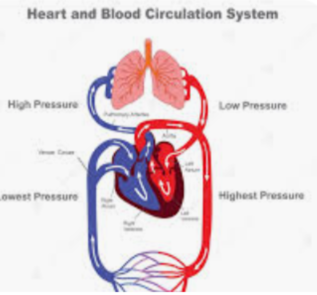
Picture taken from: ShutterStock
Blood Flow Through the Heart
Blood enters the right atrium from the vena cava. The vena cava is the largest vein, and all veins connect to it. This blood is deoxygenated and must be oxygenated to form oxyhaemoglobin.
Blood flows from the right atrium to the right ventricle through the bicuspid (mitral) valve and is transported to the lungs via the pulmonary artery.
In the lungs, blood is oxygenated and returns to the heart through the pulmonary veins into the left atrium.
Blood then moves from the left atrium to the left ventricle through the tricuspid valve. The left ventricle pumps blood through the aorta via the aortic (semilunar) valve.
The aorta distributes oxygenated blood throughout the body. Because it handles high pressure, the aorta has thick muscular walls. Veins, in contrast, have thinner walls and a larger lumen.
Heart Conditions
Congestive Heart Failure (CHF)
CHF occurs when the heart cannot pump blood effectively, usually due to conditions that overwork the heart. Excess fluid in the blood or high blood pressure forces the heart to pump faster, causing strain and death of heart tissue. This leads to fluid buildup within the heart chambers and other parts of the body.
Cardiac Arrest
Cardiac arrest is a sudden stop of the heart beating. Prior to arrest, symptoms may include shortness of breath, palpitations, and chest discomfort.
Heart Attack (Myocardial Infarction)
A heart attack occurs when heart tissue begins to die due to blocked blood and oxygen supply from the coronary arteries. Blockages are caused by plaque (fat buildup) which may fully block the artery or rupture, causing clot formation.
Symptoms include chest pain (angina), fatigue, cold sweat, nausea, lightheadedness, shortness of breath, and pain radiating to other upper body areas.
Treatments:
- Vasodilators to widen arteries and improve blood flow
- Diuretics to reduce excess fluid and lower blood pressure
- CPR or AED to stimulate heart function
- Oxygen therapy to prevent further heart tissue death
- Blood thinners to reduce clot formation
- Blood pressure-lowering medications to reduce heart strain
- Coronary angioplasty to prevent future heart attacks
Effects on the Body
- Shortness of breath due to poor oxygen transport
- Fluid buildup and swelling, particularly in legs
- Reduced blood flow to the brain can cause unconsciousness or coma
Angioplasty
Angioplasty is a procedure used to widen narrowed blood vessels caused by atherosclerosis (plaque buildup). It helps restore proper blood flow and prevent heart attacks.
Procedure Steps
- Angiogram: Medical imaging is done to locate blockages. The area is numbed beforehand.
- Catheter insertion: A needle is inserted into an artery, and a thin tube (catheter) is guided inside.
- Dye injection: Dye visible on X-ray shows the blood flow and blockage site.
- Balloon procedure: A deflated balloon on the catheter is positioned at the blockage and inflated to widen the artery.
- Stent placement (if needed): A metal tube (stent) may be inserted to keep the artery open. The balloon inflates to expand the stent, then is removed while the stent locks in place.
Angioplasty: Pros & Cons
Pros
- Less invasive than open heart surgery, making it a lower-risk option
- Costs less than a surgical procedure
- Smaller wound compared to traditional surgery
Cons
- Internal bleeding caused by over-inflation of balloon or stent
- Discomfort experienced by the patient during inflation of balloon or stent
- Arteries can become blocked again if a stent isn’t placed (restenosis)
- Possible allergic reaction to the dye used in the procedure
Blood
Red Blood Cells (Erythrocytes)
- Concave disc shape
- Smaller than white blood cells
- Main function is to transport oxyhaemoglobin
- Use passive transport to carry oxygen
- Do not contain mitochondria
- Produced in red bone marrow
- Formed in the same location as white blood cells and platelets
White Blood Cells (Leucocytes)
Phagocytes / Monocytes
- Non-specific immune attackers
- Carry out phagocytosis to engulf bacteria
- Collect at infection sites to ingest harmful microorganisms
- Kidney-shaped nucleus
Lymphocytes
- Produce specific antibodies
- Include B-cells and T-cells
- Large circular nucleus
Neutrophils
- First cells to arrive at infection sites
- Destroy microorganisms
- Cause inflammation
- Poly / star-shaped nucleus
Basophils
- Involved in allergic responses
- Release histamine and other chemicals
- Cause inflammation and allergic reactions
- Mononucleus split at the top
Eosinophils
- Fight bacteria and parasitic infections
- Bilobed nucleus
Lysosomes
Lysosomes contain proteins and enzymes that help destroy bacteria and other pathogens within immune cells.
Ventilation
Picture taken from: Theory Pages – Labster
Adaptations for Efficient Ventilation
Alveoli
- Thin walls for rapid diffusion
- Moist surface to allow gases to dissolve
- Large surface area
- Rich blood supply from capillaries
Nose
- Nasal hairs filter dust particles
- Mucus traps bacteria and pathogens
- Turbinates increase surface area for air–mucus contact
- Rich blood supply warms and humidifies air
Intercostal Muscles
- Located between the ribs
- Assist inhalation and exhalation
- Expand and contract the chest cavity
Diaphragm
- Contracts during inhalation, increasing thoracic cavity volume
- Relaxes during exhalation
- Works antagonistically with intercostal muscles
Gaseous Exchange
Gaseous exchange is a passive process. Oxygen is inhaled and diffuses into the blood, while carbon dioxide diffuses from the blood into the lungs and is exhaled.
In humans, gaseous exchange occurs in the alveoli and surrounding capillaries. In plants, oxygen diffuses out of the stomata while carbon dioxide diffuses into the plant.
digestive System
Digestion Overview
Types of Digestion
Mechanical digestion includes mastication (chewing) and peristalsis (churning food into chyme).
Chemical digestion involves enzymatic breakdown of food. Example: pepsin breaks down proteins in the stomach.
Note: Protease is activated in the stomach’s acidic environment (low pH), unlike most enzymes.
5 Phases of Digestion
- Ingestion
- Movement through esophagus
- Mechanical & chemical digestion
- Absorption
- Egestion
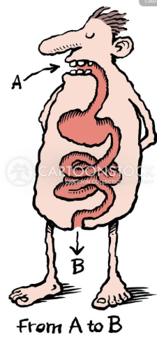
The Gastrointestinal Tract
A continuous tube connecting: mouth, esophagus, stomach, liver, gallbladder, small intestine, large intestine, and rectum.

Mouth
- Mechanical: Teeth break food, tongue mixes with saliva
- Chemical: Amylase digests starch
- Forms bolus → passes through epiglottis
- Bucal cavity = mouth region
Esophagus
- ~10 inches long
- Mucus lubricates food + traps bacteria
- Peristalsis moves bolus
- Uses antagonistic muscle pairs
Stomach
- Mechanical: Churning produces chyme
- Chemical: Enzymes + HCl (pH ~1.5)
- Protease & lipase present
- HCl kills bacteria
- Low mucus → heartburn
- Food stays ~4 hours
Small Intestine
- ~7 meters long
- Main site of absorption
- Pancreatic amylase present
- Villi & microvilli increase surface area
Large Intestine
- ~5 feet long
- Water absorption
- Bacterial digestion
- Concentrates waste → rectum
Appendix
- Larger in herbivores
- Contains bacteria to digest cellulose
Colon
- Part of the large intestine
- Absorbs water and salts from undigested food
- Forms and stores feces
- Contains bacteria that assist digestion
- Moves waste by peristalsis
- Connects the small intestine to the rectum
Small Intestine
- Longest part of the digestive system
- Consists of the duodenum, jejunum, and ileum
- Main site of digestion and nutrient absorption
- Receives bile from the liver and pancreatic juice from the pancreas
- Contains villi to increase surface area
- Absorbs nutrients into the bloodstream
duodenum
- This is the first part of the small intestine
- Receives chyme from the stomach
- Receives bile and pancreatic juice
- Bile emulsifies fats
- Pancreatic enzymes digest proteins, fats, and carbohydrates
jejunum
The jejunum is the middle section of the small intestine, located between the duodenum and ileum. It plays a major role in the absorption of nutrients.
Structure
- Approximately 2.5 meters long
- Highly folded inner surface
- Contains villi and microvilli
- Very rich blood supply
- Neutral pH
Function
- Main site of nutrient absorption
- Absorbs glucose, amino acids, and fatty acids
- Works after digestion begins in the duodenum
Adaptations
- Villi increase surface area for absorption
- Microvilli (brush border) further increase efficiency
- Thin walls allow rapid diffusion
- Dense capillary network transports nutrients quickly
ileum
The ileum is the final section of the small intestine, following the jejunum. It completes nutrient absorption and connects to the large intestine.
Structure
- Approximately 3–4 meters long
- Contains villi and microvilli
- Thinner walls than jejunum
- Fewer folds compared to jejunum
Function
- Absorbs remaining nutrients
- Absorbs bile salts for reuse
- Absorbs vitamin B12
- Passes undigested material to large intestine
Adaptations
- Villi increase surface area
- Microvilli enhance absorption efficiency
- Capillary networks transport nutrients
- Lacteals absorb fatty acids and glycerol
Special Features
- Contains Peyer’s patches (immune tissue)
- Helps defend against pathogens
- Controls flow into large intestine via ileocecal valve
Stomach
- Muscular, J-shaped organ
- Stores and churns food to form chyme
- Secretes gastric juice containing acid and enzymes
- Hydrochloric acid kills germs and aids digestion
- Pepsin begins protein digestion
- Thick mucus lining protects stomach walls
Digestive Enzymes
Amylase
- Produced in the mouth and pancreas
- Breaks down carbohydrates and starch
Lipase
- Produced in the pancreas
- Breaks down fats
Protease
- Produced in the pancreas
- Breaks down proteins
Enzyme reaction example:
2H2O2 → 2H2O + O2
Malnutrition
Kwashiorkor
- Protein deficiency disease
- Common in children in developing countries
- Swollen belly due to fluid buildup
- Thin muscles with fat-looking appearance
- Dry, scaly skin and reddish hair
- Weakness and irritability
Prevention
- Consume protein-rich foods (milk, eggs, pulses, fish, meat)
- Maintain a balanced diet
Marasmus
- Deficiency of both protein and calories
- Common in young children
- Extreme thinness and visible bones
- Sunken eyes and wrinkled skin
- Stunted growth
Prevention
- Provide balanced diet with proteins, carbohydrates, and fats
- Ensure proper infant nutrition
Respiratory Diseases
Asthma
Asthma is a chronic respiratory disease involving inflammation and narrowing of the airways.
- Triggered by dust, pollen, smoke, cold air, or exercise
- Causes wheezing, chest tightness, and coughing
Management
- Avoid triggers
- Use prescribed inhalers
- Maintain clean surroundings
Emphysema
Emphysema is a chronic lung disease that damages alveoli, reducing gas exchange efficiency.
- Mainly caused by smoking
- Causes shortness of breath and fatigue
Management
- Quit smoking
- Avoid pollution
- Use inhalers or oxygen therapy
Transpiration
Transpiration is the process of water leaving a body.
In Humans
Water vapour is released through breathing and sweating, helping regulate body temperature and remove salts.
In Plants
Water is absorbed from the soil by active transport, moves up the xylem, evaporates in mesophyll air spaces, and diffuses out through the stomata.
| Name | Purpose |
|---|---|
| Xylem | Transports water; dead tissue |
| Phloem | Transports sucrose; living tissue; bidirectional flow |
Factors Affecting Transpiration
- Humidity: Higher humidity reduces transpiration rate
- Light intensity: Increased light opens stomata
- Wind speed: Higher wind increases transpiration
- Leaf surface area: Larger area means more stomata
Measuring Transpiration
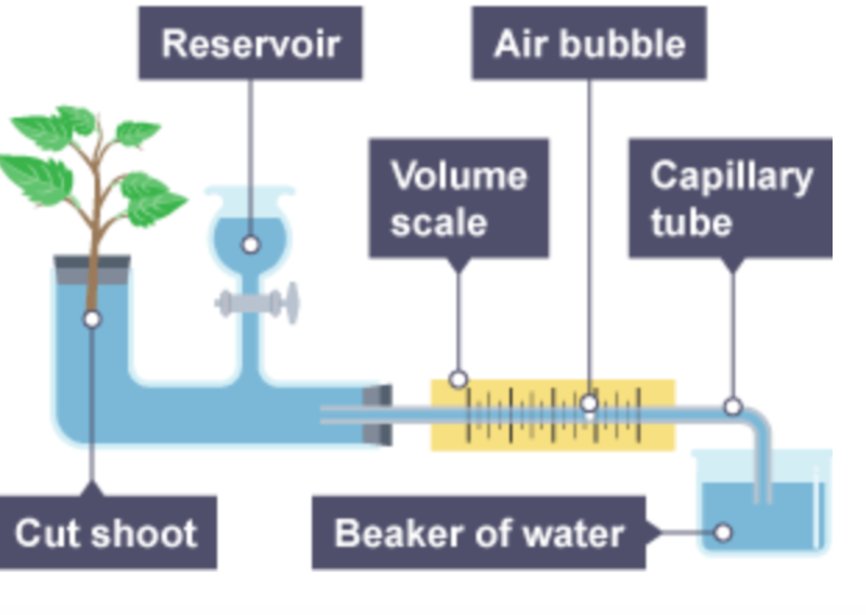
A potometer measures water uptake as a proxy for transpiration. As transpiration occurs, water is absorbed, moving an air bubble along a scale to calculate the rate.
Muscles
Found in the mesodermal layer (between the skin and the bone), muscles make up 40-50% of body mass and are:
- Excitable: Capable of receiving and responding to stimuli
- Contractile: Able to contract upon stimulation
- Extensible: Able to stretch upon stimulation
- Elastic: Capable of returning to resting position after stretching
Types of Muscle Tissue
- Skeletal Muscle Tissue: Multinucleated, rich in mitochondria, voluntary, fatigue-prone, striated, connected to the peripheral nervous system
- Smooth Muscle Tissue: Uninucleate, tapered ends, non-striated, involuntary, resistant to fatigue, found in walls of hollow organs, responsible for peristalsis and lens focusing
- Cardiac Muscle Tissue: Uninucleate, striated, involuntary, rich in mitochondria, branched, connected via intercalated disks for wave-like contraction of the heart
Muscles connect to bones through tendons, while bones connect to other bones through ligaments, both types of connective tissue.
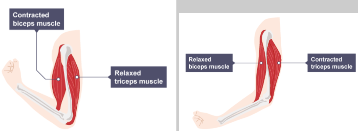
Antagonistic Muscle Pairs
Muscles can only contract and relax—they cannot push. To move bones, muscles work in antagonistic pairs:
- Agonist: The muscle that contracts → “doing all the work”
- Antagonist: The muscle that relaxes → “enjoying the agonist’s agony”
Example: When flexing the arm, the bicep contracts (agonist) while the tricep relaxes (antagonist). When extending the arm, roles reverse.
Human Skeletal System
The largest bone in the human body is the femur, while the smallest are the ossicles in the ear: the incus, malleus, and stapes (or hammer, anvil, and stirrup).
The human skeleton is an endoskeleton (located inside the body). Most vertebrates have endoskeletons, while some animals, like insects, have exoskeletons (external skeletons that are periodically shed and regrown).
Divisions of the Skeleton
-
Axial Skeleton:
- Skull
- Ossicles
- Hyoid (jawbone)
- Backbone / vertebral column
- Rib cage
-
Appendicular Skeleton:
- Arm bones
- Shoulder girdles
- Leg bones
- Pelvic girdle
Anatomy of a Bone
- Periosteum: Thin, tough membrane covering the outermost part of the bone, rich in blood vessels
- Compact Bone: The hardest part of the bone; made of living cells, protein fibers, and minerals like calcium and phosphorus
-
Spongy Bone: Found at the ends or beneath compact bone, with multiple spaces for strength without extra weight
-
Bone Marrow: Soft tissue inside the spongy bone
- Red Bone Marrow → forms red blood cells
- Yellow Bone Marrow → contains fat cells and plasma
-
Bone Marrow: Soft tissue inside the spongy bone
Joints
Ball-and-socket joints: Found in shoulders and pelvis. They allow for movement in almost all directions.
Hinge joints: Found in the knees and elbows. They facilitate one-way movement.
Pivot joints: Found in the neck, allows for side-to-side and up-and-down movement.
Gliding joints: Found in the fingers and toes (tarsals and metatarsals).
Excretory System:
Main parts of this system include the kidney, the ureter, the urethrea, the renal vein, the renal artery, the renal corpuscle and the renal pevis
Nephrons can be found in both the cortex, or outer region of the kidney, and the medullary or inner region of the kidney. These facilitate the filtration of blood and can be divided into three parts: the Malpighian body, the Proximal convoluted tubule and the distal convoluted tubule
Malpighian Body:
Glomerulus: A network of small blood vessels surrounded by Bowman’s capsule, connected to the afferent and efferent arterioles. It is where filtration of waste and excess water from blood takes place, the former two which are moved into the Bowman’s capsule.
Bowman’s capsule: A sac-like structure which surrounds the glomerulus and through which the filtered waste products pass to enter the proximal convoluted tubules. It acts like a sieve, only allowing products like water and solutes to pass through and preventing larger structures, like red blood cells, from entering the nephron. What passes through is called the filtrate.
Afferent and efferent arterioles: The afferent arterioles supply blood to the glomerulus, and the efferent arterioles transport blood from the glomerulus to the proximal convoluted tubules. These arterioles branch out and form a cage-like structure around the tubules to reabsorb what’s diffused out of the filtrate.
Renal Tubules:
These are the parts of the nephron where much of the filtered substances are reabsorbed into the body, before what is finally left is removed as urine.
Proximal Convoluted Tubules:
Sodium chloride, or salt, exits the nephron and moves back into the fluid surrounding the nephron to be reabsorbed back into the bloodstream. Water diffuses out through osmosis and is also eventually reabsorbed by the body. Glucose and amino acids are also filtered out here. The twisting structure of the PCT provides more time for all necessary substances to be reabsorbed back into the bloodstream. Drugs and toxins are secreted into the nephron here.
Loop of Henle:
The descending limb of the Loop of Henle is permeable to water but not sodium, causing a buildup of sodium as the filtrate travels down. However, the ascending limb of the loop is permeable to sodium but not water, causing the sodium to diffuse from an area of high concentration to low concentration and exit the nephron. Water and sodium are filtered out here as well.
This creates a gradient where the area at the bottom of the loop is “saltier” while that at the top is more diluted, or “watery”.
Reverse Osmosis:
Reverse osmosis is a separation technique where pressure is applied to a solution to force the solvent to move from an area of low concentration to one of high concentration, leaving behind the heavier solute particles.
It is called reverse osmosis because it goes against the natural process of osmosis, where solvent moves from an area of high concentration to one of low concentration.
This occurs in Bowman’s capsule in the kidneys when filtering waste from the blood. The afferent arteriole is larger than the efferent arteriole, which creates a region of high pressure in the glomerulus. This high pressure goes against osmotic pressure and forces solutes out of the solvent, resulting in reverse osmosis and the filtration of waste and excess fluid from the blood.
Dialysis:
Dialysis is the process where a machine called a dialyser performs the role of a kidney when the latter is unable to function properly. It removes toxins from the blood and filters them out when kidneys cannot do so. It is also used to remove drugs from the body.
One type of dialysis is called hemodialysis, where blood is filtered and cleaned outside the body before being supplied back in.
Procedure:
- To access both oxygenated and deoxygenated blood for filtration, an arteriovenous graft or fistula is created. The former is done by connecting an artery and a vein with a small plastic tube called a graft. In the case of the latter, they are connected directly surgically.
- Two needles are inserted into the graft or fistula, which are connected to tubes that connect to the dialyser.
- The impure blood from a person enters a dialyser, where it is passed through filtering fibres to filter out the waste and is mixed with a liquid called a dialysate. These filtering fibres are filled with the dialysate, and are tube-like in nature. These tubes serve as a semipermeable membrane, through which only water, electrolytes, and toxins are allowed to pass through into the dialysate through diffusion and osmosis.
- Certain medicines and drugs may be added into the dialysate to diffuse into the patient’s filtered blood, such as erythropoietin, which is typically produced by the kidneys.
- The cleaned blood is pumped back into the body, while the dirty dialysate fluid is pumped out of the machine. Clean dialysate fluid is constantly supplied back into the machine.
Pros:
- Flexibility: Hemodialysis can be done in a hospital setting or at home.
Cons:
- Dietary restrictions: Patients undergoing hemodialysis are recommended to avoid consuming excess fluids and certain foods.
- Restrictive: Hemodialysis needs to be regularly conducted for an average of three days a week, each session lasting four hours. This takes up a lot of the patient’s time.
- Increased risk of sepsis (blood poisoning): Due to the insertion of an external catheter into the bloodstream, there is risk of contamination and the entry of bacteria into the blood if proper sanitation is not ensured. Sepsis can be fatal.
A second type of dialysis is called peritoneal dialysis, where blood is filtered and cleaned within the body itself, particularly within the membranes of the abdomen called the peritoneum.
Peritoneal Dialysis:
Procedure:
- A catheter, or a small, flexible tube, is inserted into the abdominal membranes and fills the abdominal cavity with dialysate.
- The dialysate draws out harmful substances and excess fluids from the body.
- The used dialysate is filtered out of the body and into a disposable bag via the catheter.
Pros:
- Can be done at home due to not requiring being constantly hooked up to a dialysis machine, unlike hemodialysis.
Cons:
- Peritonitis, or the infection of the abdominal membrane: Due to an external device like the catheter being repeatedly inserted and removed into the peritoneum, there is a risk of contamination and infection.
- Weight gain: The dialysate fluid contains sugar molecules, which can be absorbed back into the body and increase caloric intake as a result. Without compensating for this by exercising or reducing caloric intake, this can lead to eventual weight gain.
Organ Transplantation:
What?
A medical procedure where a damaged organ or tissue in a patient, called the recipient, is removed and replaced with a healthy, functioning one from an organ donor.
Successful transplants have been conducted for multiple organs and body tissues, save for the brain and a few others. Some, like the kidneys and liver, are easier to transplant than others. The average human being is born with two kidneys (not me though, I have three), but can live normally even with only a single functioning kidney. As a result, kidney donations are extremely accessible and easy to set up.
Livers, on the other hand, everyone only gets one of. But the neat thing is that parts of the liver can regenerate, meaning that chunks of a person’s liver can be donated to a recipient with liver failure, with the donor recovering their original liver in a short span of time.
Kidney and liver organ transplants are some of the most common organ transplants worldwide as a result.
When?
These are usually conducted in life-threatening situations, such as genetic conditions like cystic fibrosis or being born with an organ defect.
Whom?
Organ donors:
- Must consent prior to registering themselves as donors.
- Must be 18 years or older if donating organs while alive, such as kidneys and liver.
- Must be brain dead if donating organs posthumously.
- Must have a compatible blood type. If blood types between donor and recipient are not compatible, then the recipient’s immune system may view the donor’s blood cells as antigens and generate an immune response, causing the former’s body to reject the donated organ. People with an O- blood type are universal donors and can donate to anyone, but can only receive from other O- blood type holders.
- Must be healthy. Though for some transplants, health isn’t a requirement, when donating organs like the liver, one must not be suffering from any conditions associated with the organ, as they would risk transferring the disease to the recipient.
Why?
The benefits of organ donation include:
- Donating organs means saving lives. In the U.S. alone, more than 103,000 people are waiting for organ donations, with almost 17 dying each day waiting to receive one.
- Donating your body to science allows medical schools and students to learn about human anatomy and physiology through hands-on experience.
…But?
Organ donations can go wrong in several ways, such as:
- Not receiving organs on time: Certain organs can only survive outside the body for a short period of time, meaning that if the surgery is done too late, the donation as a whole is rendered useless.
- Weakened immune system due to immunosuppressants: Immunosuppressants are drugs given to organ recipients prior to the surgery where they receive their new organ to weaken the immune system and prevent it from generating an immune response. This is due to the fact that the immune system may view the foreign organ as a harmful antigen and may attack it, causing the recipient’s body to reject the organ and not integrate it into the body. However, a weakened immune system results in the patient being more susceptible to infections by pathogens while recovering from the transplant.
- Religious beliefs preventing organ donation: Certain religions may frown upon and altogether ban those practicing them from being organ donors, reducing the availability of donors further.
- Social pressure: Organ donors may feel pressured by friends, family, or loved ones to whom they may be donating an organ, resulting in a case of dubious consent.
- Organ trafficking: The practice of removing organs illegally from dead or living persons, often regardless of whether consent was given or not, and selling said organs, which is another illegal act.
However…
Newer, modern alternatives to organ transplants are emerging. One example is 3D organ printing, or bioprinting.
What?
This technology uses real cells in combination with 3d printers to essentially “3d print” new organs and artificially create them. This can either be done by directly 3d-printing pieces of tissue and organs, or by 3d-printing a mold or structure of said organ in which cells can be injected to replicate.
The cells in question can be taken from donors or from the patient themselves.
Homeostasis
What is homeostasis?
Homeostasis is the maintenance of internal physical and chemical conditions within a normal range in response to external or environmental changes or stimuli. The nervous system and the endocrine system are both responsible for homeostasis.
Homeostasis is something we can only do because we are endotherms, a.k.a warm-blooded animals that can control our own body temperature. Well, us mammals along with the birds. Reptiles and amphibians, a.k.a “cold-blooded” animals, are ectotherms, whose body temperature changes based on their surroundings and is out of their control. That’s why reptiles spend so much time basking in the sun, to warm themselves up.
The three key components of homeostasis, which together make up the body’s control system responsible for maintaining homeostasis, are:
Receptors:
These are cells or structures responsible for sensing stimuli and relaying the sensation to the coordination centres. They can be classified based on the location of the stimuli they receive as either exteroceptors or interoceptors.
| Exteroceptors | Interoceptors |
|---|---|
| Detect and are responsive to external stimuli like a change in external temperature, touch, or pain. | Detect and are responsive to stimuli from internal organs and tissues inside the body, like glucose levels or blood pressure. |
| Usually located near the surface of the body like in the skin. | Usually located inside or near organs themselves. |
| Thermoreceptors (sense temperature), photoreceptors (detect light), mechanoreceptors (respond to pressure or touch). | Baroreceptors (detect blood pressure), nociceptors (detect pain), chemoreceptors (detects chemical changes within the body like pH levels or oxygen levels in the blood). |
They can also be classified based on the type of stimulus they receive:
| Steady-state receptors | Changing-state receptors |
|---|---|
| Generate impulses as long as a certain state, such as temperature or pressure, is maintained. | Generate impulses only if the intensity or position of a stimulus changes. |
Coordination centres:
These organs are responsible for receiving the stimulus sent from the receptors and sending an appropriate input to the effectors to respond to the stimulus. The brain and spinal cord are the typical coordination centres.
Effectors:
These organs receive inputs from the coordination systems and respond to the stimulus accordingly. These are typically muscles or glands.
Ex:- Blood sugar levels:
Receptors: Liver.
The liver detects either a rise or fall in blood sugar levels beyond the normal range and sends a stimulus to the pancreas.
Coordination centres: Pancreas.
The pancreas receives the input from the liver and acts accordingly.
Effectors: Pancreas.
In the case of a blood sugar crash, the pancreas secretes glucagon, an enzyme which signals to the liver to break down stored glycogen into glucose to enter the bloodstream.
In the case of a blood sugar spike, the pancreas secretes insulin, an enzyme which signals to the liver to store excess glucose in the bloodstream as glycogen.
To summarize:
To connect the receptors, coordination centers, and effectors, the nervous and endocrine system both come into play.
homeostasis is generally made at:
- A body temperature of around 37 degrees Celsius/96.5 degrees Fahrenheit.
- A blood pH of around 7.3.
- A blood sugar level between 80-110 milligrams/decilitre.
- A body made up of 70% water, and blood that is 90% water by volume.
- And finally, enough carbon dioxide, but not too much! Otherwise, cells will begin to oxidize. This creates oxidative stress, which makes your cells quite literally break down.
The Nervous System:
Neurons:
Neurons, or nerve cells, are of three types:
| Sensory Neuron | Interneuron/Relay Neuron | Motor Neuron |
|---|---|---|
| Transmits information as electrical impulses from receptors that detect stimuli to the CNS. | Transmits information between sensory neurons, motor neurons, and the CNS. | Transmits information as electrical impulses from the CNS to effectors, which are either muscles or glands. |
Reaction Time:
Reaction time is the time between the introduction of a stimulus and the start of a reflex, and is a measure of how readily an individual responds to a stimulus. It can be measured through the simple falling ruler experiment done with yourself and a friend.
- Hold the ruler by the end so that the “zero” marking is aligned with the thumb and index finger of the other person.
- Release the ruler.
- Using a stopwatch, measure the time taken for your friend to catch the ruler.
- Using the final position of your friend’s fingers, calculate the distance covered by the ruler while falling till it was caught.
- Divide the distance by the time, and you get the reaction time in meters per second!
Reaction time can be influenced by factors such as alcohol or drugs, age, type of stimulus, distractions, and fatigue.
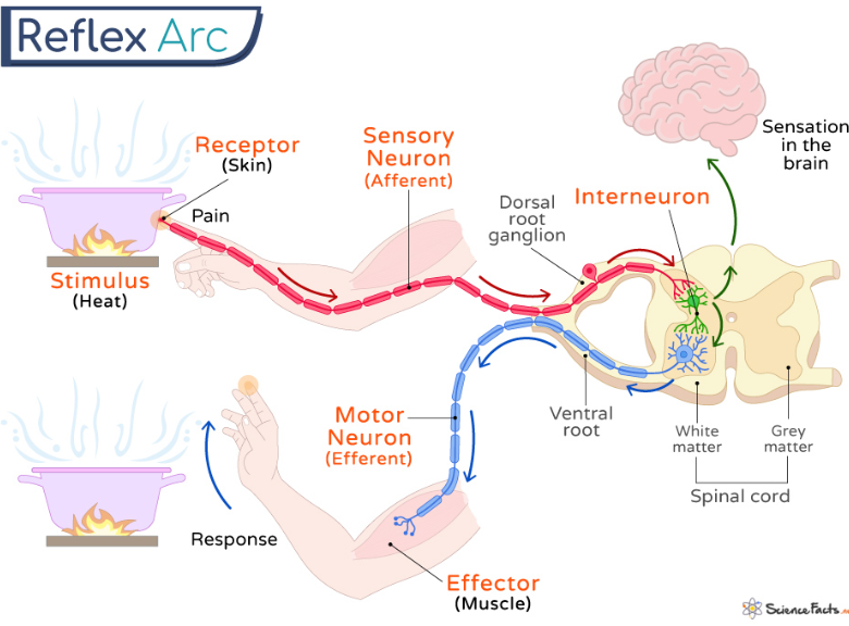
Reactions are voluntary, while reflexes are involuntary. Reflex arcs are pathways followed by nerve impulses in reaction to a stimulus. An example of a reflex is the knee-jerk reflex. Voluntary actions are controlled by the brain, while involuntary actions are controlled by the spinal cord, which serves as a bridge between receptors and the brain, and the brain and effectors as well.
Don’t Be So Impulse(ive)!
A nerve impulse is an electrical signal that propagates through neurons to convey a message or signal in the nervous system. They can travel up to 120 meters per second through nerve fibres!
…Nerve fibres?
Well, you didn’t think it just stopped at nerves, didn’t you?
Nerves themselves
Nerves themselves are made of anywhere between just a few to a million nerve fibres, also known as fascicles, all bundled together by a covering called the perineurium. The fibres themselves have a width between 5-20 micrometers, as wide as the blood capillaries in your body!
Structure:
As far as cells go, they’re pretty simple. Nothing more than a cell membrane with cytoplasm inside. However, this “simple” structure is the backbone for their functioning.
Function:
Within both the cytoplasm inside the membrane and the fluid outside, there are positively and negatively charged ions. However, their distribution in these respective regions is imbalanced. And an imbalance of charges creates a potential difference, also known as voltage! Now, typically, this voltage stays at -70 mV.
However, when an impulse occurs, channels in the membrane open. These allow sodium ions from the outside to diffuse inside, causing the voltage to rise to +40 mV. Then, different channels allow potassium ions from the inside to diffuse outside, causing the voltage to fall back down to -70 mV.
Finally, pumps in the membrane move the sodium and potassium ions to their initial positions in preparation for another response.
Nerve impulses are…
- All or Nothing: When a nerve impulse occurs, the voltage either rises all the way to 40 mV, or it doesn’t occur at all.
- One-Way: Nerve impulses can’t go back the way they came. They can only move in one direction.
- Domino Effect: Adding on to the fact that they can’t go back, nerve impulses can’t travel over the gaps called synapses. Instead, neurotransmitters bridge the gap and trigger an impulse in the next nerve fibre, and so on and so forth, creating a domino effect.
…Neurotransmitters?
These are chemicals that are released at synapses to bridge the gap between neurons and stimulate an impulse in the next neuron, because charges can’t cross the gap! There are many types of neurotransmitters, but they can roughly be divided into excitatory and inhibitory neurotransmitters.
Excitatory neurotransmitters cause the next nerve cell’s voltage within the membrane to rise, triggering an impulse! They “excite” the neuron, hence their name. Too much of this, however, called overexcitation, can cause panic attacks and anxiety, like if there’s too much norepinephrine in your head, which excessively triggers the fight-or-flight response.
Inhibitory neurotransmitters cause the next neuron’s voltage to drop even further, meaning an even larger amount of excitatory neurotransmitter is required to stimulate an impulse. They “inhibit” impulses, hence their name. Once again, though, if there’s too much of this, sedation can occur, making you feel sleepy and inactive.
Reaction time
The interval between the presentation of a stimulus (visual, auditory, or tactile) and the initiation of a voluntary response is what reaction time is defined as. It is measures the speed of nervous system processing, involving perception, mental processing, and motor execution.
Factors influencing reaction time:
Effect: Moderate amounts decrease reaction time (faster responses).
Scientific Reasoning: It blocks adenosine receptors in the brain, preventing drowsiness and increasing neuron firing rate, which enhances alertness and speeds responses.
Effect: Stronger stimuli reduce reaction time.
Scientific Reasoning: Stimulus intensity such as brighter lights or louder sounds activate sensory receptors more strongly, generating larger nerve impulses that reach the brain more quickly.
Effect: Practice decreases reaction time.
Scientific Reasoning: repeated actions strengthen neural pathways and increase myelination(Formation of myelin sheath around nerve fibres), allowing impulses to travel faster.
Effect: Children and older adults usually have slower reaction times than young adults.
Scientific Reasoning: Young adults generally have the fastest reaction times. In children, the nervous system is still developing, while in older adults nerve conduction velocity and synaptic efficiency decline.
Effect: Increase reaction time.
Scientific Reasoning: The brain must divide attention and filter irrelevant stimuli, increasing processing time before responding.
Effect: Less sleep increases reaction time (slower responses).
Scientific Reasoning: Sleep deprivation reduces cognitive function and alertness because of disruption in neurotransmitter balance, delaying signal transmission across synapses.
Effect: Extremely cold slows reaction time; moderate warmth can improve it.
Scientific Reasoning: Enzyme activity and ion movement in neurons decrease, reducing nerve impulse speed.
Effect: Usually increase reaction time.
Scientific Reasoning: It slows down central nervous system activity and synaptic transmission, delaying perception and response.
Effect: Dominant hand usually reacts faster.
Scientific Reasoning: Neural pathways controlling the dominant hand are more frequently used and more efficiently wired, leading to faster motor output signals.
Effect: Mild stress may decrease reaction time; extreme stress increases it.
Scientific Reasoning: Mild stress can decrease reaction time due to adrenaline increasing alertness, but excessive stress increases reaction time because cortisol impairs concentration and decision-making.
Effect: Low glucose increases reaction time.
Scientific Reasoning: The brain relies on glucose for ATP production needed for active transport in neurons.
Effect: Dehydration slows reaction time.
Scientific Reasoning: Electrolyte imbalance interferes with nerve impulse conduction and reduces blood flow to the brain
Brain Anatomy
The brain and spinal cord make up the central nervous system, while the nerves that connect the brain and spinal cord to the other parts of the body are part of the peripheral nervous system.
The brain itself can be divided into the cerebrum, which is the largest part, the thalamus, and the hypothalamus. The cerebrum is divided into two hemispheres: the left and right hemispheres, which control opposite sides of the body. The left controls the right side of the body, and vice-versa.
The cerebrum is divided into four lobes: the frontal lobe, parietal lobe, occipital lobe, and temporal lobe.
The frontal lobe is responsible for thinking, including planning, problem solving, and speech.
The parietal lobe is responsible for regulating and processing sensory inputs from receptors.
The occipital lobe is in charge of visual stimuli and sight.
The temporal lobe works with the ears to process auditory stimuli.
The cerebellum, which is located beneath the cerebrum, is responsible for voluntary actions and maintaining the body’s balance and equilibrium. Finally, the brainstem controls involuntary actions.
Positive and Negative Feedback Loops
Negative feedback loops are more common and involve the reversal of the original stimulus. These occur when bodily conditions change from the ideal range. They typically involve the process of decreasing then increasing levels to stay within the normal range. This can be observed throughout a variety of bodily processes, such as:
Blood Pressure
Changes in blood pressure are picked up by baroreceptors in the walls of the aorta and carotid arteries, which supply blood to the brain. These detect the stretch of the walls in response to blood volume per unit of time.
If blood pressure is low, impulses are sent to the brain, which triggers the release of aldosterone and antidiuretic hormone (ADH) from the kidneys. Aldosterone signals the kidneys to retain sodium, causing water retention as well due to osmosis. ADH signals the kidneys to release less urine. The increased fluid and narrowing of blood vessels (vasoconstriction) help raise blood pressure.
If blood pressure rises, blood vessels dilate (vasodilation) and heart rate decreases to lower it.
Body Temperature
External conditions can cause body temperature to drop or rise. Thermoreceptors sense these changes and relay the information to the brain, which compares it to the body’s normal temperature range.
If temperature is too low, vasoconstriction occurs to prevent heat loss, and shivering happens, which is rapid muscle contraction and relaxation producing heat. Body hairs stand on end (goosebumps) to trap heat.
If temperature rises, vasodilation occurs, and sweating helps cool the body via evaporation. Sweat glands, which originate from the epidermis and extend into the dermis, produce sweat made of water, salt, and dissolved urea.
Water Levels (Osmoregulation)
Changes in internal water levels are picked up by osmoreceptors, which relay this information via impulses to the hypothalamus. The hypothalamus stimulates the posterior pituitary gland to release ADH (AntiDiuretic Hormone), which affects how permeable the renal tubules in the kidneys are to water.
If the body is dehydrated, the cells in the collecting ducts become more permeable, allowing water to diffuse back into the bloodstream. More ADH is secreted to conserve water. More water is reabsorbed back into the blood, and the urine produced is more concentrated, lower in volume, and darker yellow in color.
If the body has excess water, the collecting duct cells become less permeable, causing most water to be flushed out as urine. Less water is reabsorbed, and less ADH is released. Urine is larger in volume, more dilute, and lighter in color or even colorless.
The formal term for internal water levels is blood water potential, which measures how much water is in the blood. Too much can cause high blood pressure; too little leads to dehydration.
Positive Feedback Loops
Positive feedback loops are less common and involve the amplification of the original stimulus. They encourage continued changes in the same direction and can be harmful if conditions exceed equilibrium, but are useful in certain processes.
Blood Clotting
Blood clots form through a series of enzyme activations. One enzyme, thrombin, activates both the next and previous enzymes in the sequence, creating a positive feedback loop. Thrombin levels continue to rise until the clot is fully formed.
Childbirth
During labour, the baby’s head stretches the cervix. This stimulus causes the hypothalamus to signal the pituitary gland to release oxytocin, which causes uterine contractions. Contractions further stretch the cervix, creating a positive feedback loop until the baby is born.
In this process:
Receptor: cervix, senses the stimulus of the baby’s head.
Control center: brain/pituitary gland, produces oxytocin.
Effector: uterus, contracts and widens the pelvic region.
Oxytocin levels continue rising until after delivery, then reset to normal.
Endocrine System
Anterior Pituitary Gland
The anterior pituitary gland secretes several important hormones:
- Adrenocorticotropic hormone (ACTH): Stimulates the adrenal cortex to release cortisol.
- Follicle stimulating hormone (FSH): Stimulates egg development in ovarian follicles in females and triggers ovulation, and stimulates sperm cell development in testes of males. FSH release is controlled by GnRH from the hypothalamus.
- Luteinizing hormone (LH): Triggers sex hormone release in both males and females. In females, signals ovaries to produce estrogen and progesterone, and release a mature egg. LH release is controlled by GnRH from the hypothalamus.
Posterior Pituitary Gland
The posterior pituitary gland does not synthesize hormones but stores those produced by the hypothalamus:
- Antidiuretic hormone (ADH): Stimulates kidneys to reabsorb water when the body is dehydrated, resulting in more concentrated urine. Alcohol inhibits ADH, leading to diluted urine and dehydration.
- Oxytocin: Released during physical labour, such as childbirth.
Pineal Gland
A small gland in the brain that secretes melatonin, regulating the sleep cycle by reducing heart rate and other body processes. High light intensity detected by photoreceptors inhibits melatonin, while low light encourages its production.
Thyroid Gland
Located at the base of the neck. It secretes:
- Thyroxine: Regulates metabolism of carbohydrates, fats, and proteins. Iodine deficiency can cause goitre (swelling at the base of the neck).
- Calcitonin: Reduces calcium levels in the blood, counteracting parathyroid hormone.
Parathyroid Glands
Located next to the thyroid gland. They secrete parathyroid hormone (PTH), which increases calcium levels and counteracts calcitonin.
Thymus
Produces thymosin, which stimulates production of T-cells, a type of white blood cell.
Adrenal Glands
Two glands located on top of the kidneys. They secrete adrenaline and cortisone.
Adrenaline Effects:
- Increases heart rate, boosting oxygen supply to skeletal muscles.
- Stimulates diaphragm and intercostal muscles to enhance gas exchange in lungs.
- Diverts blood to skeletal muscles and away from digestive system and skin.
- Stimulates glycogenolysis, breaking down glycogen into glucose.
The adrenal cortex (largest part) secretes:
- Aldosterone: Regulates blood pressure by increasing sodium and fluid retention.
- Cortisol: Released during stress, part of the fight-or-flight response, increases blood glucose.
- Androgens: Trigger puberty and development of male and female sex characteristics.
Adrenal Medulla
The adrenal medulla is the inner part of the adrenal gland. It secretes:
- Adrenaline (epinephrine)
- Noradrenaline (norepinephrine): Released from both the adrenal gland and the brain, it functions as both a hormone and a neurotransmitter. Works with adrenaline in the fight-or-flight response by:
- Constraining blood vessels to increase blood pressure
- Increasing blood glucose levels
- Enhancing attention and focus
Pancreas
The pancreas regulates blood glucose levels via specialized cells:
- Beta cells: Secrete insulin, which lowers blood sugar by signaling the liver to store glucose as glycogen. A deficiency in insulin leads to diabetes.
- Alpha cells: Secrete glucagon, which raises blood sugar by signaling the liver to release stored glucose.
Ovaries
These are typically only found in those with XX chromosomes, or females. They produce oestrogen, which is responsible for the development of secondary sexual characteristics in females during puberty. During the menstrual cycle, oestrogen thickens the lining of the uterine walls to prepare for ovulation. It also triggers the release of luteinizing hormone (LH).
Testes
These are sex organs typically only found in those with XY chromosomes, or males. They produce testosterone, which is responsible for the development of secondary sexual characteristics in males during puberty, as well as triggering sperm production.
Plant Hormones and Behaviours
Plant movements can either be tropic (relating to the growth of the plant) or nastic (irrelevant to the growth of the plant).
Nastic movements include:
- The touch-me-not plant shutting its leaves when receiving the stimulus of being touched.
- The Venus flytrap shutting its leaves when an insect lands on them and gets trapped.
Tropic movements can either be hydrotropic (movement towards water), phototrophic (movement towards light) or geotropic (movement towards soil).
As plants lack a nervous system, they rely on hormones for regulating growth and other activities. However
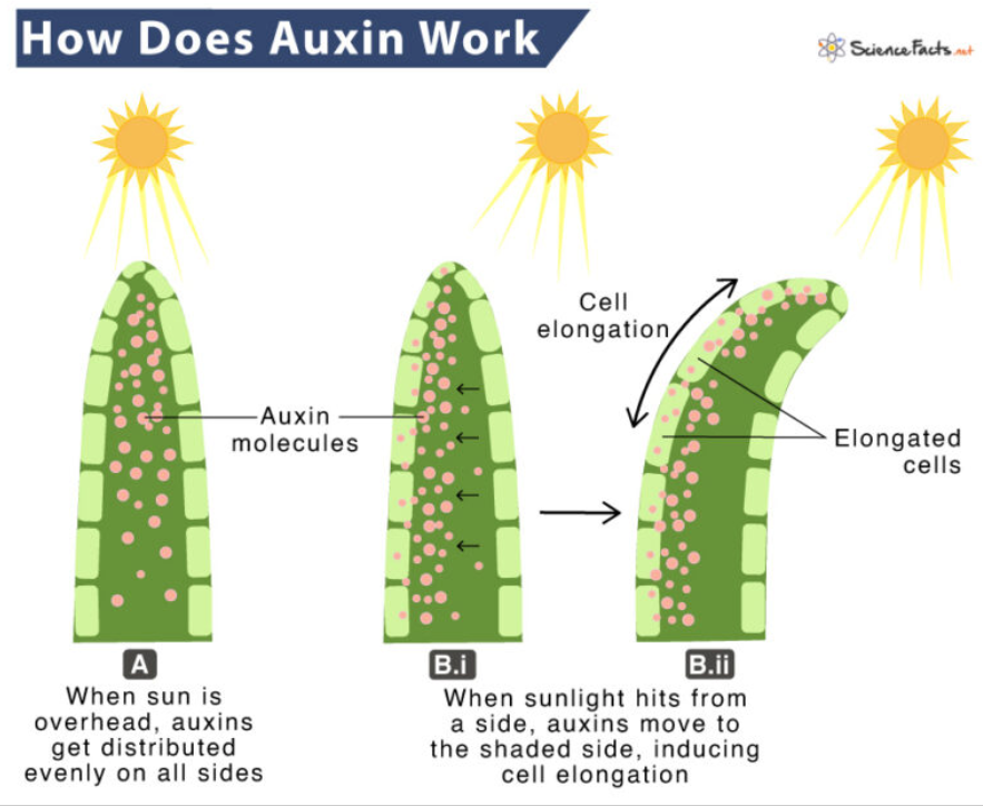
Auxins in the shoots
Auxins avoid light. Thus, when exposed to light, auxins migrate to the part of the shoot that is in the shade. As a result, the part of the shoot in the shade begins to grow at a faster rate than the part of the shoot exposed to the light. This causes the plant to bend towards the light.
Auxins are also greatly influenced by gravity. In a shoot growing horizontally, auxins will accumulate at the lower side, causing it to grow faster. As a result, the plant curves upwards.
Auxins in the roots
In the roots, auxins inhibit growth. As roots grow out, gravity causes auxins to become concentrated on the bottom side of the root. Thus, the bottom side of the root grows slower than the upper side, which grows down due to gravity. Thus, roots exhibit geotropism.
Commercial uses of auxin
- Weed killer/herbicide: Too much of a good thing can be a bad thing, and that is especially true for auxins. Excess auxin within a plant stimulates the release of the enzyme ethylene, which inhibits the growth of roots and shoots and makes the plant die.
- Furthermore, auxin only affects dicotyledons, and not monocots or grasses. Thus, farmers who grow monocot crops like rice spray herbicide containing auxins in their fields. This causes any dicot weeds to grow too fast and die, while allowing the desirable monocot crops to continue growing.
- However, once sprayed in a field, the spread of auxins can’t be controlled, and may negatively impact the surrounding environment if it leaches out.
- Cloning plants: Farmers can take a cutting of a desirable plant and dip the tip of the cutting in a solution of auxins, which causes the rapid development of roots and the simple creation of clones. A similar process is used in laboratory settings.
Gibberellin
This is a plant hormone responsible for two main functions within a plant: stem elongation and seed germination.
Stem elongation:
Gibberellins are produced near the nodes of where new leaves are growing. They are secreted at a low concentration to encourage cells in the stem of the plant to elongate. As a result, the plant increases in height.
Plants with a lack of gibberellin are considered dwarf plants, while those with excess amounts grow very tall.
Furthermore, they also promote the elongation and overall increase in size of fruit, as well as the flowering of plants.
Seed germination:
Gibberellins trigger the hydrolysis of starch in the endosperm of the seed into glucose, which allows the plant embryo to begin germination.
Commercial uses of gibberellin:
Aesthetic purposes: Gibberellin can be harvested as a solution that is then sprayed on plants to increase growth, promote flowering, and increase the size of fruits.
Cytokinins
Coming from the word “cytokinesis”, which is a stage in cell division where the cytoplasm is split in two as the two daughter cells are produced, these hormones aid in cell division.
Abscisic acid
Plant hormones that regulate the plant’s response to stress, and act throughout the plant in a variety of ways:
Seeds:
A buildup of abscisic acid in the seed of a plant prevents the seed from germinating and prolongs its dormant phase. This typically occurs when the seed is in unfavourable conditions to germinate. However, once conditions become less harsh, abscisic acid finally ceases to inhibit gibberellins and allows seed dormancy to end.
Roots:
If the plant detects low water levels in the surrounding soil, the roots produce abscisic acid. This stimulates the elongation of roots and increases the root’s ability to conduct water through itself. It also inhibits lateral root growth and focuses only on making roots grow deeper.
Leaves:
Due to low water levels, abscisic acid from the roots travels to the leaves, and forces water out of guard cells through reverse osmosis, causing them to shrivel up and close the stomata, preventing further water loss.
Furthermore, during autumn, abscisic acid causes trees to shed their leaves. As temperatures drop, groundwater in the soil becomes frozen, preventing the tree from utilizing it. Thus, it is unable to replace the water lost through transpiration, hence it sheds its leaves.
Furthermore, leaves are unable to support the weight of snow in winter, resulting in the breaking of branches that have a larger load of snow piled on top. Thus, leaves are shed.
Buds:
Abscisic acid is produced in undeveloped, or terminal flower buds. This causes the buds to enter a period of dormancy and not flower until conditions are favorable. It also results in the conversion of the leaves that accompany the bud into bud scales, which are harder and protect the bud against mechanical stress.
Pavlov and Classical Conditioning
Ivan Pavlov was a Russian scientist whose discovery of the psychological phenomenon of classical conditioning arose from his experiment with dogs. He observed that the dogs would salivate when they were given food, as well as in the presence of the lab technician who usually gave them food.
Intrigued, he designed an experiment where he would ring a bell while presenting the dogs with food, and measure their saliva output. While initially, there was no response to the bell alone, the pairing of the bell with the food led the dogs to associate the noise with salivation.
Pavlov began to observe that the dogs produced a similar output of saliva when he began to ring the bell alone. The main principle behind Pavlov’s experiment is this: a stimulus that triggers a natural biological response is paired with a new stimulus that triggers the same reaction.
Unconditioned Stimulus (Food)
The food was an unconditioned stimulus, which is a stimulus that triggers a natural biological response. In this case, the stimulus of the food activates receptors in the eyes (rods and cones) and the nose (olfactory receptors), which send electrical impulses to the control centre, the brain. The brain interprets the stimulus and sends impulses to the effectors, in this case the salivary glands, which execute the natural biological response of salivation.
The natural behavioural response of salivation when presented with food is called an unconditioned stimulus, because it occurs naturally.
Unconditioned stimulus → Unconditioned response
Food → Salivation
Neutral Stimulus (Bell)
The sound of the bell ringing was initially a neutral stimulus. This means that while the stimulus was picked up by the dog through auditory receptors in the ears and was relayed to the control centre via electrical impulses, it did not trigger any natural biological responses.
Neutral stimulus → No response
Bell ringing → No response
Conditioning Process
Over time, as the bell was rung along with giving food to the dogs, synaptic connections were formed and strengthened between the neurons that relayed the auditory stimulus to the brain, and the neurons that stimulated the salivary glands to release saliva.
Food → Salivation
Bell ringing → No response
Food + Bell ringing → Salivation
Eventually, the bell ringing became a conditioned stimulus, meaning through external conditioning it now mimics an unconditioned stimulus and triggers a conditioned biological response.
Conditioned Stimulus and Conditioned Response
Neutral stimulus → Conditioned stimulus
The dogs then began to salivate at the sound of the bell ringing alone, due to the formation of a neural pathway where the auditory stimulus relayed to the brain via electrical impulses resulted in the brain sending signals to the effectors (salivary glands) to release saliva.
This makes salivation a conditioned response, as it is a natural biological response that now occurs in response to a conditioned stimulus.
Conditioned stimulus → Conditioned response
Bell ringing → Salivation
Extinction
But this isn’t a cheat code for infinite dog saliva– not that you should want infinite dog saliva in the first place– because of extinction.
If a conditioned stimulus is repeatedly presented without the unconditioned stimulus that triggers the same response, eventually, the association between the conditioned stimulus and the conditioned response will begin to fade.
Bell ringing (🛇 Food) x 10 → No salivation
Meaning that if the bell is rung in front of the dog enough times without giving it food, then the dog will stop associating the noise with salivating. This is called extinction.
Limits of Classical Conditioning
Furthermore, classical conditioning can only trigger pre-existing natural biological responses with new stimuli, and can’t create new behavior. So Pavlov could make his dogs drool when he rang a bell, but he couldn’t make them do backflips.
Additionally, the conditioned response is not an exact replica of the unconditioned response. Pavlov found that the chemical composition of the saliva released when he rang the bell was slightly different from the saliva released at the sight of food alone.
Brief Notes on Eye, Tongue, and Ear Anatomy
The eye, ears, and tongue are all sense organs, which are specialized structures in the human body that allow the individual to receive and respond to specific types of stimuli in the environment.
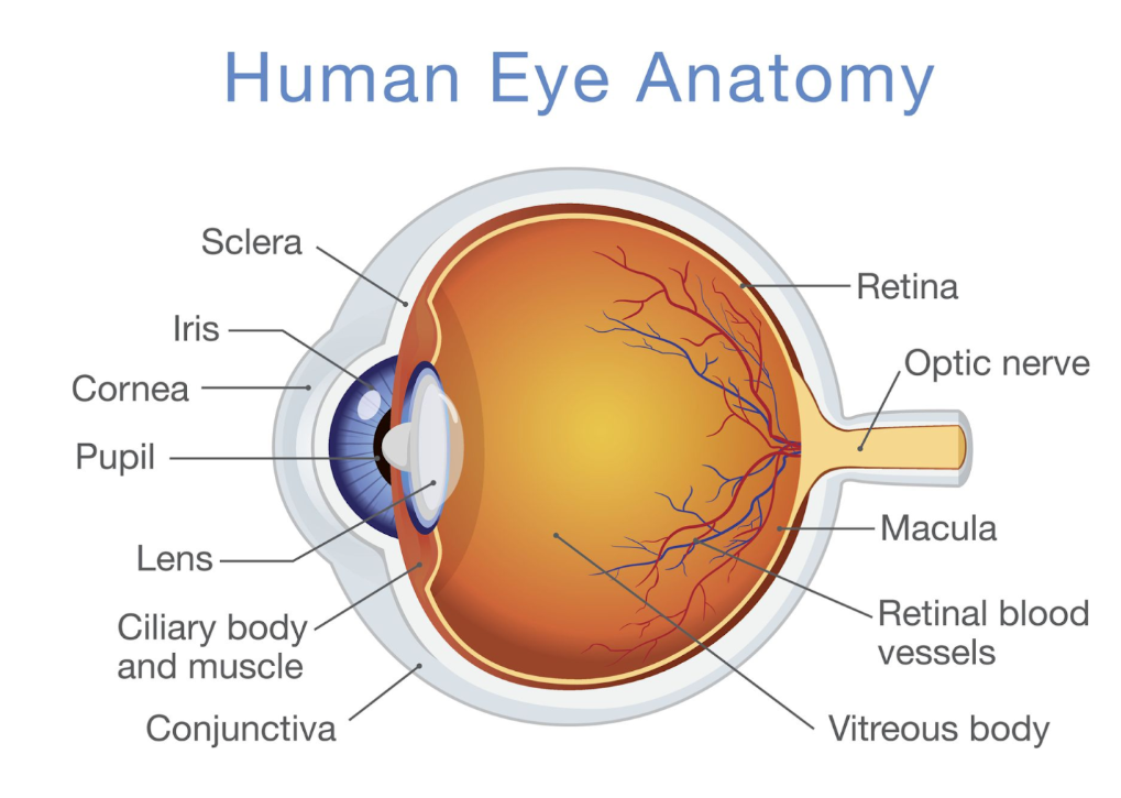
The Eye
This sense organ is responsible for vision and visual stimuli. The parts of the eye are as follows:
Cornea: A transparent, curved structure located in the front of the eye. It allows light to enter the eye, and refracts all light entering the eye so it is focused on the lens and retina. It also protects the iris, pupil, and lens.
Iris: A colored structure located behind the cornea. It is responsible for controlling the amount of light entering the eye by adjusting the size of the pupil. The colour of the iris also determines eye colour.
The iris reflex is the involuntary contraction and relaxation of the iris in response to a light stimulus. If the eye is exposed to high-intensity lighting, the iris contracts and constricts the pupil to reduce the amount of light entering the eye and prevent damage to the retina. If the eye is exposed to low-intensity lighting, the iris relaxes and dilates the pupil to increase the amount of light entering the eye which allows the eye to see better in dim lighting.
Pupil: An aperture through which light enters the eye whose size is controlled by the iris. There are also two sets of muscles in the pupil, the circular muscles and the radial muscles. These are smooth muscles, meaning the action they perform is involuntary! I mean, when was the last time you saw someone control their pupils?
To dilate the pupils, the radial muscles contract while the circular muscles relax. To constrict the pupils, the circular muscles contract while radial muscles relax. Thus, these guys also form an agonist-antagonist muscle pair!
Ciliary bodies: Muscles that control the width and thickness of the lens by contracting and relaxing, regulating the degree to which light entering the eye is refracted.
Aqueous humour: A transparent fluid in the eye located between the iris and the cornea. It exerts pressure on the eye, supplies nutrients, and refracts light.
Lens: A transparent, biconcave structure which refracts light entering the eye. Its thickness and width are controlled by the ciliary bodies.
Vitreous humour: Transparent fluid in the eye located behind the lens and in front of the retina. It exerts pressure on the eye to maintain its shape and delivers nutrients to the retina.
Retina and fovea: The retina is a thin layer of light-sensitive tissue at the back of the eye. It contains photoreceptors, which are types of receptors sensitive to visible light.
Photoreceptors: There are two types of photoreceptors: the rods and the cones. Rods are sensitive to dim lighting conditions and help in night vision. However, they are not sensitive to colored light, which is why everything appears grey in dim lighting or darkness. Cones are sensitive to high-intensity lighting and colored light, but are useless at night.
The fovea is a point in the retina which has a large concentration of cones. Light that is projected onto the fovea creates the sharpest image.
Optic nerve and the “blind spot”: The optic nerve is a bundle of nerve cells that transmits electrical impulses from the photoreceptors to the brain where they are interpreted as images. It also regulates the iris reflex.
The blind spot is where the optic nerve cuts into the retina where there are no photoreceptors. Any light that is reflected onto the blind spot is not picked up by photoreceptors, so the brain automatically fills in the image in the blind spot based on information from the surroundings.
The movement of light into the eye is as follows:
Light → Cornea → Aqueous humour → Pupil → Lens → Vitreous humour → Retina → Fovea → Photoreceptors → Optic Nerve → Brain
The Ear
This sense organ is responsible for auditory stimuli, or hearing, but some parts of the inner ear also regulate body balance.
The anatomy of the ear is as follows:
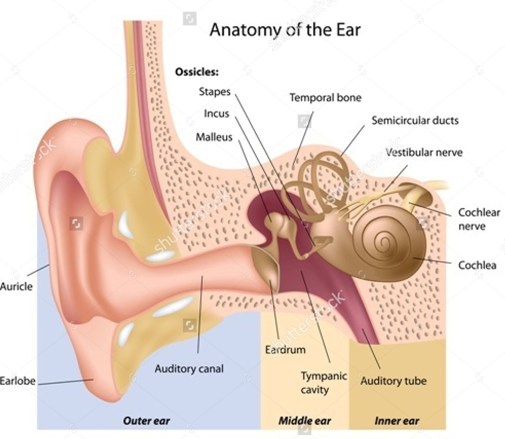
The ear is divided into three parts: the outer ear, the middle ear, and the inner ear.
Outer ear:
Pinna: A structure made of cartilage that funnels sound waves into the auditory canal towards the middle and inner ear.
Middle ear:
Eardrum: A thin, sensitive membrane connected to the ossicles that vibrates when sound waves hit it.
Ossicles: The smallest bones in the body that are connected to the eardrum and vibrate along with it as sound waves travel through the ear. The vibration of the ossicles amplifies sound waves as they enter the inner ear.
Inner ear:
Cochlea and cochlear nerve: The vibrations from the ossicles cause the fluid in the cochlea to ripple, which is picked up by sensitive hairs in the cochlea that send electrical impulses to the brain via the cochlear nerve, where they are interpreted.
Semicircular ducts: These tube-like structures contain fluid that moves when the head or body changes position. The fluid movement is picked up by sensitive hairs, which relay this through electrical impulses to the brain.
The Tongue
This sense organ is responsible for picking up on taste stimuli from food. It contains tiny projections on the surface called papillae (singular: papilla) that contain taste buds connected to nerve fibres that send electrical impulses to the brain.
Ossicles: The smallest bones in the body that are connected to the eardrum and vibrate along with it as sound waves travel through the ear. The vibration of the ossicles amplifies sound waves as they enter the inner ear.
Inner ear:
Cochlea and cochlear nerve: The vibrations from the ossicles cause the fluid in the cochlea to ripple, which is picked up by sensitive hairs in the cochlea that send electrical impulses to the brain via the cochlear nerve, where they are interpreted.
Semicircular ducts: These tube-like structures contain fluid that moves when the head or body changes position. The fluid movement is picked up by sensitive hairs, which relay this through electrical impulses to the brain.
The Tongue: This sense organ is responsible for picking up on taste stimuli from food. It contains tiny projections on the surface called papillae (singular: papilla) that contain taste buds connected to nerve fibres that send electrical impulses to the brain.
There are different types of papillae found on the tongue:
- Filiform papillae: Most numerous type; provide texture and grip for food but do not contain taste buds.
- Fungiform papillae: Mushroom-shaped structures found mainly on the tip and sides of the tongue; contain taste buds.
- Circumvallate papillae: Large papillae arranged in a V-shape at the back of the tongue; contain many taste buds and are sensitive to taste.
- Foliate papillae: Located on the sides of the tongue; contain taste buds and are more developed in children than adults.
Criteria B and C:
Hypothesis: If the [independent variable] is increased/decreased, the [dependent variable] will increase/decrease because [scientific explanation]
Research question: How does the [independent variable] affect the [dependent variable]
Identifying variables: The variables are often categorized based on how they are changed
Independent variable: is being changed
Dependent variable: Changes as a result of the independent variable changing– is being measured
Control variables: Does not change at all
When writing variable of an experiment, there must be 1 independent variable, 1 dependent variable and 2 (or more) control variables
Manipulation of variables:
This means how the independent variable is being changed, how the dependent variable is measured and how the control variable is being kept constant. Write one paragraph for each.
→ Most of this information is already given to you in the question so don’t try to do anything fancy or use your knowledge just write what is given, remember to read the question carefully.
Method: When writing any safe and logical method, ensure to list all the materials, describe safety precautions and then list all the steps. If safety measures and/or materials required are needed in another question skip this and only write the method/procedure
Materials: xyz(amount), abc (amount).......
Safety measures: the researcher will (safety caution, for example; wearing gloves) because (why the safety caution is necessary, for example avoiding contact with the skin because the thing is corrosive). Write two safety measures, you can write more but it is not necessary
Method:
xyz
abc
def
mno
qrs
Adding an improvement and extension to the experiment:
This one is extremely easy after you have identified how you’re measuring your dependent variable and what your independent variable is.
Making an extension to the experiment means changing the independent variable, don’t change it to something incredibly off topic, say, if the experiment is on photosynthesis and the independent variable was leaf surface area; you can change this to other factors affecting photosynthesis, like light and carbon dioxide. If you’re feeling fancy and know more about this topic you can write about auxins and photoreceptors but that is seldom required (unless you want your teacher to like you more).
Writing an improvement to the experiment is how to increase accuracy, the most failproof improvement is conducting more trials and using a more precise measuring tool. There’s normally potential areas of improvement throughout the question so remember to read everything carefully.
Validating the hypothesis:
Write the hypothesis in past tense, backing up the result using data collected
“ When the (independent variable) was increased/decreased, the (dependent variable) increased/decreased because (mini data analysis)”
Validating the method:
2 strengths of the method and 2 weaknesses– four lines minimum.
Graph:
This is a 5 mark question, based on if the student includes all the below required points:
X-axis title
Y-axis title
Scale of x and y axis (written like: x axis– 1 unit= x (unit measurement), y axis– 1 unit= x (unit measurement)
Graph title (just summarise the research question)
Accurate data interpretation (draw each line to the correct value change)
If you have all of these and they are correct you simply cannot lose marks. Remember that the x-axis is the independent variable and the y-axis is the dependent variable.
Criteria B and C (continued)
“Explain a research question”:
This means you have to state the research question(s) of the experiment and break it down. Add on to what the method is and how it works, discuss what is being changed, what is being measured and what is being controlled and how this benefits the investigation.
Example:
Explain the Research question.
The research question is:
“How does gravity affect the direction of root growth in seedlings?”
This question aims to investigate whether gravity influences the angle at which plant roots grow.By comparing seedlings grown under normal gravity (Petri dish placed on its side) with seedlings grown on a rotating clinostat(where the effect of gravity is reduced), students can determine if gravity is responsible for directing root growth downward.
Keeping the seedlings in darkness ensures that light does not affect the results, so only gravity is being tested.
Graph analysis:
This is where you interpret what each trend shown on the graph means, using points of the graph to validate the scientific explanation and whether it was correct or incorrect.
Remember to include the graph type (like bar or line graph) , why this graph type is most suitable and how the trend shown by the graph validates the scientific reasoning.
Criteria D:
In most cases this will be an essay or an evaluation task.
How to write a good scientific essay:
Any good iB biology essay will have an introduction to the problem, an evaluation of the solution, how the topic shows impacts socially, economically,environmentally, culturally or technologically- pick any 3 and describe it in detail with justifications. After this you will conclude your essay through highlighting what you’ve found from your evaluation. It follows the following format:
(there will be some background information provided, the essay will almost always be about the solution to a problem)
Introduction :
set the problem in context, touch on what it is. Then set the solution in context while touching on some strengths and limitations of this solution. Remember to use scientific terminology in this part and throughout your response
Procedure:
Describe the procedure of the problem. How does it start? Is it contagious? How does it escalate? How long does the escalation take? Is it treatable? If so, how? Relate the procedure of the problem to terminology used in the specific science– for example if it was on angioplasty use terminologies such as fatty plaque, pacemaker and cellular respiration.
Impacts (environmental/economic/social/cultural/technological/political):
This will be 3 paragraphs, each talking about one impact. For each, try writing 3 positive and 3 negative impacts with justification
Environmental:
How does the solution impact the environment? This is the easiest to connect anything to because either the solution will require more advanced technologies (therefore making it environmentally unsustainable because of land cleared and energy used for production) or it will be holistic and require less technology, making it more environmentally sustainable because no toxic chemicals are released, no land is cleared for production and little to no power source is required.
Provide solutions to negative environmental impact with appropriate justification and scientific reasoning (you kind of gotta use your brain for this because you need to come up with and come up with a solution for each impact)
Social:
You can relate this by saying how the problem affected an individual’s work productivity along with caregiver burden then explaining how the solution wolves this to an extent.
Later going into depth about how the solution shows social impacts with proper justification. Does it help with mobility? Does it improve productivity? Does it minimise caregiver burden?If the thing has a negative social impact, suggest solutions to that.
Economical:
This one is incredibly easy to connect most solutions to, this is because they often require more technology and therefore need more finances to function. This makes it economically unsustainable for the middle and lower classes because they are seldom able to afford proper medical treatment.
You can even suggest solutions to this such as government funding or donation drives.
Technological:
This is kind of basic and you can include points on how advancements in technology are used throughout the solution and how they may improve the solution. You just discuss the overall technological impact this solution has.
Political:
It’s quite annoying that most people do not know how to connect sciences to politics so please practise this. You can draw connections such as how wars lead to different access to technology. For example, in the cold war period communists would not have access to the same technology as capitalists and vice versa right? It is because of a country’s political relationships that they have access to a certain technology or solution to a problem.
Cultural:
Most cultures uphold holistic strategies to diseases, you can compare these strategies with medically developed strategies to connect your topic to culture.
Conclusion:
Revise your introduction with the scientific evaluations you made throughout the essay. This should be the same length as your intro.
Here’s an exemplar 8/8 essay from a ninth grade past paper about antibiotic resistance and immunization:
To what extent has the development of medicinal sciences promoted societal health and well being ?
As medicinal sciences have developed in order to most efficiently counteract harmful miccroorganisms through antibiotics over recent decades, pleomorphic properties of pathogens have permitted for a phenomenon known as antibiotic resitance,whhere they mutate in a waay thaat allows them to survive antibiotic treaaatments. This introduces economical as well as social issues as researchers must constantly invent new antibiotics to counteract these evolving micrroorgaanisms. Moroever, this raises ethical debates as the cost of effective medical treatment increased, therefore financiallly challenges or underdeveloped communities would not have equal access to the correct disease treatment, creating questions revolving around to what extent are antibiotics able to promote social wellbeing; where social wellbeing may be defined as a state of physical health,disease risk and work productivity.
What is an antibiotic?
Firstly, an antibiotic is a scientifically designed tool used in order to counteract harmful microorganisms from infecting an individual. Throughout history, society has used similar anti-microbials to treat infectious disease, however developments into what is now known as antibiotics showed most significance in the 1900s— with the invention of a compound known as Salvarsan and the discovery of penincillin. Antibiotics work by entering a targeted microorganism, binding to specific sites and inhibiting vital pprocesses. Taking the example of penincillin; Penincillin enters a bacterial micro organism and blocks cell wall synthesis— resulting in lysis and the cell’s osmotic prressure becomes too high.
What is antibiotic resistance?
However, with the development of antibiotics and their widespread usage throughout society— microorganisms have begun to develop what is known as ‘Antibiotic Resistance’. Antibiotic resistance is where the pathogen mutates in a way that allows them to survive antibiotic treatment. The highest level of antibiotic resistance is otherwise known as pleomorphism, and is most commonly seen in viral infections— where the antibiotic is rendered useless as the microorganism has completely mutated in a way that is resistant to the same. Over the recent decades, antibiotic resistance has been recognised as an increasingly troubling issue as the increased societal access to antibiotics has led to higher levels of mutation towards antibiotic resistance in microorganisms. In order to combat this, the development of new antibiotics is required; however this creates economic and environmental impacts as well as raises social and ethical debates.
What is the impact of an antibiotic on human health and social wellbeing?
The usage of antibiotics yields significant social impacts as it is able to prevent individuals from disease on the large scale. This can promote decreased chance of pandemic and increased productivity as more healthy individuals are able to work efficiently. However, as the microorganism evolves to survive in the conditions of antibiotic treatment, the usage of antibiotics can often yield higher negative than positive impacts on the individual’s health. This is because as the pathogen develops antibiotic resistance, treatment is rendered useless or yields an insignificant positive impact on the disease; however, the individual would continue to face the financial burden of antibiotic treatment plans, alongside decreased work productivity and health as the disease progresses.
What is the environmental impact of antibiotic usage?
Moreover, the usage of antibiotics is argued to show a significant impact on the environment. The usage of antibiotics in fields such as agriculture have permitted for extended animal life as risk of disease is reduced significantly. However, the creation of antibiotics and required scientific research demands higher energy expenditure alongside large areas of cleared land. This creates environmental arguments as it is a major contributor to the emission of carbon dioxide gas. This is because high energy requirements are often met by the burning of fossil fuels, expanding society’s growing carbon footprint and significantly contributing to the worsening issue of global warming. Moreover, large areas of land for research institutes where scientific development into antibiotics occurs correlate with a decline of forest cover, adding on to the environmental issue of habitat destruction.
Economic impacts of antibiotics
From an economic standpoint, antibiotics show both positive and negative impacts. Firstly, the development of antibiotics has permitted expanding work productivity on a large scale. This is associated with a higher Gross Domestic Product (GDP) alongside an increased Human Development Index (HDI). Moreover, the introduction of antibiotics into agriculture has allowed for reduced disease in animals and therefore increased output in the animal-based agriculture industry.
However, antibiotics have undeniably created negative economic impacts. This is because antibiotic resistance has forced scientists to constantly create new antibiotics to counteract evolved microorganisms. This creates a financial burden for researchers, as they must continuously invest time and money into developing new drugs to combat a constantly evolving pathogen.
Ethical impacts
Moreover, whilst antibiotics have undoubtedly promoted social wellbeing, this is to a debated extent as the widespread usage of antibiotics to combat microorganisms has raised ethical controversies. This is because financially challenged areas have limited access to antibiotics which are able to counteract evolved microorganisms, creating unequal disease risk.
Moreover, ethics becomes a major debate with factors based on human error such as over-prescription, which can promote a significant decline in an individual's mental and physical health and is more likely to occur in underdeveloped communities where access to qualified medical professionals is limited.
Conclusion appraisal
In conclusion. whilst the development of antibiotics has showed signficiant positive social and economic impacts, through promoting work productivity, social well being and expanded potential in the agricultural industry— the widespread usage of antibiotics has often been debated in ththe context of ethics and environmental consequences. This is because as access to antibiotics has expanded over the recent decades, chances of over-prescription has increased espceially in udnderdeveloped communities— contributing to a decline in theh idnividuals health. Additionally, as antibiotic resistance becomes an increasingly worrying issue the consistent development of new antibiodies to comabt the evolved microorganisms is necessary, increasing the financial burden pof producing, designing and purchasing essential antibiotics alongside increasing energy and land requirements for medicinal research facilities . This rraises economic as well and environmental iiseus as it is correlated with higher costs alongside increased carbon emmisions, contributing toi societu’s cqarbon footprint. Moving forward, the sustainable development and implementation of antibiotic use through correct prescritions,, equal access to healthcare and environmental consiousness is imperative for a developed society.
Note: Most of the time when you have to write a 1000 word essay like this, people will get intimidated by the word count. This note is to tell you not to do that, the essay will naturally be over 1000 words with all of the content; the word count is just an estimate of how many words the essay will naturally come up to. Another thing is spelling, when typing in ninth grade spelling can become a major problem, but don’t waste all your exam time on this and not the content. Focus on the content first, then fix all of the spelling errors. It helps if you plan a time allotment for each part of your exam; this can be giving 45 minutes to your essay, minimum 30 minutes in criteria A and 40 minutes in B & C , giving you 10 minutes of extra time in your EOT which you can use to review the paper.
Note: You may have noticed a lot of typos throughout the essay (my keybaord was tweaking out on me lowkey)- so please keep in mind spelling is nowhere near as important as the content. Stop taking so much time to fix typos while writing the essay cause you'll just lose marks, instead if you find more time after finishing everything fix the typos then
To evaluate a source/scientific method:
Consider strengths and weaknesses, using proper scientific justification throughout. End it with an appraisal. That's all